What is breast imaging and what is it used to diagnose?
Breast imaging uses a wide variety of tools and technologies to screen, detect, and diagnose breast cancer. If cancer is detected, these tests help find the type of cancer, as well as determine the stage and location of the cancer.
Mammography
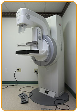
A mammogram is an X-ray image of your breasts. It can be used either for breast cancer screening or for diagnostic purposes, such as to investigate symptoms or abnormal findings on another imaging test.
Mammograms play a key role in breast cancer screening. They can detect breast cancer before it causes signs and symptoms.
Heritage Valley Health System’s board-certified mammography technologists pay special attention to optimal technique, body positioning, and breast compression to minimize your exposure to radiation during the mammogram.
Screening mammography is a routine preventative measure recommended once a year for women aged 40 and above.
Diagnostic mammography evaluates a specific problem such as an abnormality on a recent screening mammogram or a new patient concern such as pain or a lump.
3-D mammography also known as digital breast tomosynthesis is the newest and most advanced technology in mammography and allows for improved early cancer detection.
Breast Ultrasound
Breast ultrasound is an imaging technique that uses sound waves to create images of breast tissue. It is used for a closer look at findings seen on a mammogram as well as patient concerns such as lumps. Ultrasound can demonstrate whether a mass is a solid mass or a cyst.
Heritage Valley Health System breast sonographers have special training in performing breast ultrasounds to achieve the most accurate images.
Breast ultrasound is a standard, painless additional test to evaluate areas of concern found on a mammogram or breast exam.
MRI
For women and men with a breast cancer diagnosis, and for those at high risk of breast cancer, magnetic resonance imaging or MRI can provide the clearest, most detailed pictures of the breast. This radiation-free imaging technology creates 3-D images of the breasts that we use in screening, staging, treatment response evaluation, and pre-surgery planning.
This test typically lasts around 45 minutes and requires you to lie still for best results. Heritage Valley Health System’s board certified technologists provide comfort measures that include eye covers, warm blankets, and noise-cancelling headphones.
Image-guided breast biopsy
A breast biopsy is a minimally invasive procedure in which we remove a sample of breast tissue from your breast. A pathologist examines the tissue under a microscope to check for cancerous cells. We use imaging technology to precisely pinpoint the biopsy area and to guide the needle to the same area. The size, shape, and location of the tumor determine if the exam will be performed using ultrasound, stereotactic or MRI guidance.
Stereotactic Breast Biopsy
Stereotactic breast biopsy is a procedure that uses computer technology to guide a needle to an abnormality seen on mammography.
ABUS Ultrasound
ABUS ultrasound is a technology that assists in evaluating dense breast tissue as a compliment to mammography.
RSL or Radioactive Seed Localization
RSL is a small radioactive marker placed into the breast under imaging guidance by the radiologist that is detected by the surgeon in the operating room. This allows precise localization of breast abnormalities and can be done up to 5 days prior to surgery.
Risks and limitations of mammograms include:
- Mammograms expose you to low-dose radiation. The dose is very low, though, and for most, the benefits of regular mammograms outweigh the risks posed by this amount of radiation.
- Having a mammogram may lead to additional testing. If something unexpected is detected on your mammogram, you may need additional tests. These might include ultrasound, and a procedure called a biopsy to remove a sample of breast tissue for laboratory testing. However, most findings detected on mammograms aren't cancer.
- If your mammogram detects something unusual, the radiologist who interprets the images will want to compare it with previous mammograms. If you have had mammograms performed elsewhere, you will be asked for your permission to request them from your previous health care providers.
- Screening mammography can't detect all cancers. Some cancers may not be seen on the mammogram. A cancer may not be seen if it's too small or if it is located in an area that is difficult to view by mammography, such as your armpit or if your breasts are extremely dense.

